

1. Tendon structure and mechanobiology
In sport, the tendon is not a passive cable. It is a viscoelastic, mechanosensitive tissue that sits in series with muscle and determines how muscle force is transmitted, stored and released. Every sprint, jump, landing, deceleration and change of direction depends on this muscle-tendon interaction.
Structurally, tendon is dominated by type I collagen organised in a hierarchy: collagen molecules assemble into fibrils, fibrils into fibres, fibres into fascicles, and fascicles into the whole tendon, all embedded in a proteoglycan-rich extracellular matrix (Screen et al., 2015; Zhang et al., 2022). This architecture gives the tendon high tensile strength while still allowing controlled elongation under load. For training purposes, the relevant properties are mechanical rather than anatomical. Stiffness describes force per unit elongation and depends on both tendon size and material. Young's modulus describes stress per unit strain and reflects the quality of the material independent of size. Strain is the relative elongation of the tendon under load, and hysteresis is the proportion of stored energy lost as heat during unloading.
Tendon is also a living tissue that responds to strain. Tenocytes sense deformation of the surrounding matrix and convert it into collagen gene expression and protein synthesis (Kjaer, 2004). After a loading session, collagen synthesis peaks at around 24 hours and remains elevated for approximately three days, while collagen degradation also increases but peaks earlier (Magnusson et al., 2010). The first days after heavy loading are therefore a remodelling window rather than a period of immediate strengthening, which has direct consequences for where heavy tendon loading is placed in the training week (Section 5).
A further constraint is that the adult tendon core turns over very slowly. Carbon-14 bomb-pulse dating showed that the core of the human Achilles tendon is formed largely during growth and is essentially not renewed in adulthood (Heinemeier et al., 2013). This does not mean that adult tendons cannot adapt. It means that much of their training adaptation is expressed as changes in material properties rather than rapid replacement of the collagen core, which is consistent with training studies in which Young's modulus improves within weeks, well before cross-sectional area changes (Geremia et al., 2018).
From a sport-science perspective, these properties matter for three reasons.
Force transmission. A stiffer tendon transmits muscle force to the skeleton with less elongation and less delay, which favours rapid force expression during jumping, sprinting and re-acceleration.
Energy storage and return. During landing, braking and ground contact in running, the tendon stores elastic strain energy and returns part of it during push-off. Tendon compliance also allows muscle fascicles to work at more favourable lengths and velocities, which improves the efficiency of the stretch-shortening cycle (Komi, 2000; Lichtwark & Wilson, 2007).
Load tolerance. A tendon that has adapted to repeated high-force exposure is better prepared for the mechanical demands of sport. A tendon without adequate adaptation may become reactive when exposed to sudden increases in volume or match congestion, in line with the continuum model of load-induced tendinopathy (Cook & Purdam, 2009).
The training goal is therefore not maximal stiffness for its own sake. It is a tendon whose mechanical profile matches the task: stiff enough to transmit force quickly, compliant enough to store and return energy, and robust enough to tolerate repeated high-strain exposure.
2. Mechanical properties and task loading
Tendon load cannot be described adequately by naming the exercise or sport action. The most direct and widely reported descriptor is peak tendon force, the maximum force the tendon experiences during the task, normalised to body weight (BW). Peak force reflects the peak strain imposed on the tendon, which is the variable most closely linked to tendon adaptation (Section 4), and it allows sport actions and gym exercises to be compared on the same scale.
Baxter et al. (2021) applied this approach when developing an Achilles tendon loading progression for rehabilitation. Rather than ranking exercises by difficulty or appearance, they estimated the Achilles tendon force each exercise produced. Single-leg lateral and forward hopping produced the highest peak Achilles forces (7.3 BW), followed by single-leg hopping in place (6.7 BW), while running stance (5.2 BW) and two-leg hopping (4.8 BW) were lower. Moving from two legs to one, or adding a lateral or forward component, therefore raises tendon force substantially, which is why two actions that look similar on video can produce very different tendon demands.
The same logic applies to the patellar tendon. Garcia et al. (2022) showed that forward jump landings produced higher patellar tendon force than vertical jump landings (approximately 5.0 vs 4.0 BW, inverse-dynamic estimates), demonstrating that horizontal momentum increases knee-extensor demand. Verheul et al. (2024) showed that maximal horizontal deceleration from sprinting exposed the patellar tendon to peak internal forces of approximately 7.42 ± 1.84 BW and the Achilles tendon to 5.58 ± 1.80 BW, the highest patellar tendon force in Table 1. These tendon forces are comparable to those reported for high-speed running, but horizontal ground reaction forces during deceleration can reach up to six times those of accelerated or constant-speed running, with large eccentric demands on the quadriceps, soleus, tibialis anterior and gluteal muscles (Verheul et al., 2024). This is consistent with the view that decelerations are among the most mechanically demanding and potentially damaging actions in team sport (Harper & Kiely, 2018).
On smaller screens, each row is shown as a labelled block.
Study | Task | Tendon | Peak force (BW) |
|---|---|---|---|
Verheul et al., 2024 | Max horizontal decel. from sprinting | Patellar | 7.42 ± 1.84 |
Verheul et al., 2024 | Max horizontal decel. from sprinting | Achilles | 5.58 ± 1.80 |
Garcia et al., 2022 | Forward jump landing | Patellar | 5.0 ± 0.8 |
Garcia et al., 2022 | Vertical jump landing | Patellar | 4.0 ± 0.6 |
Baxter et al., 2021 | Running stance | Achilles | 5.2 ± 0.9 |
Baxter et al., 2021 | Two-leg hopping | Achilles | 4.8 ± 1.8 |
Baxter et al., 2021 | One-leg hopping | Achilles | 6.7 ± 1.6 |
Baxter et al., 2021 | One-leg lateral hopping | Achilles | 7.3 ± 2.4 |
Baxter et al., 2021 | One-leg forward hopping | Achilles | 7.3 ± 1.9 |
Values are mean ± SD. BW = body weight. Garcia values are inverse-dynamic estimates.
Read together, the table shows that tendon force depends more on the direction and constraints of the task than on its appearance. Horizontal deceleration produces the highest patellar tendon force, single-leg lateral and forward hops produce the highest Achilles tendon force, and forward or decelerative tasks consistently exceed their vertical or in-place equivalents. Common sport actions expose the patellar and Achilles tendons to roughly 5 to 7.5 BW, which defines the force range that preparation must ultimately allow the athlete to tolerate.
Two practical points follow. First, the highest tendon forces in team sport occur during single-leg, reactive and decelerative tasks, not during typical gym exercises. Second, the tasks that produce the highest forces are not necessarily the most effective adaptive stimulus. In a 14-week intervention matched for strain magnitude and volume, loading the Achilles tendon for 3 s per repetition at approximately 6.5% strain increased stiffness by 57% and Young's modulus by 51%, whereas a protocol based on fast one-legged jumps produced only non-significant trends in stiffness (Bohm et al., 2014). Sport-like reactive loading is the transfer target, but it does not replace the sustained high-strain loading that drives structural adaptation.
Tendon preparation must therefore progress through both stages: controlled high-strain loading to build capacity, then progressive exposure to the actual loading profile of the sport, including braking, landing, hopping, sprinting and change of direction, to build tolerance.
3. When Load Exceeds Capacity
The patellar tendon connects the quadriceps to the tibia via the patella. In sport, it is loaded whenever the athlete produces or absorbs high knee-extensor force: jumping, landing, squatting, braking, cutting and re-accelerating.
Patellar tendinopathy is common in jumping and multidirectional sports not because the tendon is inherently fragile, but because it is repeatedly exposed to high mechanical demand. When that demand exceeds the current load-bearing capacity of the tendon-quadriceps system, the response to loading shifts from adaptation towards accumulated matrix damage, with microrupture and material fatigue proposed as possible mechanisms (Magnusson et al., 2010).
The mechanical data are consistent with this explanation. Forward jump landings produce higher patellar tendon force than vertical landings (Garcia et al., 2022), and maximal horizontal deceleration generates peak patellar tendon forces of approximately 7.42 ± 1.84 BW (Verheul et al., 2024). The patellar tendon is therefore heavily loaded not only during jumping but also during braking and change of direction. By contrast, evidence linking specific landing kinematics to patellar tendinopathy remains limited and inconsistent (Tayfur et al., 2022), which suggests that exposure and capacity matter at least as much as technique.
At the tissue level, patellar tendinopathy involves changes in tendon structure and mechanical behaviour. Wiesinger et al. (2020) showed that athletes with proximal patellar tendinopathy present with lower tendon stiffness, stress and Young's modulus, while tendon strain, hysteresis and energy-storage capacity were preserved. Millar et al. (2021) describe tendinopathy as involving changes in tendon matrix, collagen organisation, cellular activity, pain processing and function.
Patellar tendinopathy is best understood as a load-capacity problem rather than a single-cause injury. Load-side factors include jump and landing volume, sprint and braking exposure, change-of-direction frequency and approach speed, match congestion and insufficient recovery, surface changes, acute load spikes and poor load monitoring. Capacity-side factors include the structural properties of the patellar tendon, quadriceps strength and eccentric braking ability, tendon stiffness and material properties, load tolerance and training history, and pain sensitivity and quadriceps inhibition.
Pain also changes how the athlete moves and how the nervous system drives the quadriceps. Rio et al. (2015) showed that athletes with patellar tendinopathy present with reduced quadriceps output and cortical inhibition, and that heavy isometric loading can acutely reduce both pain and inhibition. Patellar tendinopathy is therefore not only a local tissue problem. It also disrupts neuromuscular function at the knee, which is why the neural effects of loading, discussed in Section 4, are clinically relevant.
4. Loading methods: Isometrics, Eccentrics and Sport Transfer
The loading stimulus: magnitude, strain and duration
One of the most consistently supported findings in tendon adaptation research is that load magnitude matters. Bohm et al. (2015), in a systematic review and meta-analysis, concluded that tendon adaptation is strongly influenced by loading magnitude. The tendon does not adapt simply because a contraction is isometric, eccentric or concentric. It adapts when the mechanical stimulus is sufficient. Arampatzis et al. (2007) demonstrated this directly: high-strain cyclic loading of the Achilles tendon increased stiffness, elastic modulus and regional cross-sectional area, whereas low-strain loading of similar volume did not.
The physiological driver is tendon strain, with a window of roughly 4.5 to 6.5% associated with structural change. Percent MVC is a practical surrogate rather than the mechanism, and the relationship between intensity and strain varies between individuals. In practice, a threshold above approximately 70% MVC appears practical for achieving high tendon strain in most healthy individuals, although depending on the individual the target strain may be reached anywhere between about 30% and 90% MVC (Merry et al., 2022). Strain duration also matters: about 3 s of loading per repetition produced larger gains in stiffness and modulus than either 12 s loading or high-rate jumping at matched strain and volume (Bohm et al., 2014).
The time course of adaptation is faster than often assumed. During 12 weeks of high-load plantar flexor training, Achilles tendon stiffness increased by 51% and Young's modulus by 87% within the first 4 weeks, cross-sectional area changed only from week 8 (+15%), and after 12 weeks stiffness had increased by 82%, modulus by 86% and cross-sectional area by 17% (Geremia et al., 2018). Early gains are therefore mainly material, and later gains add a structural component.
Isometric loading
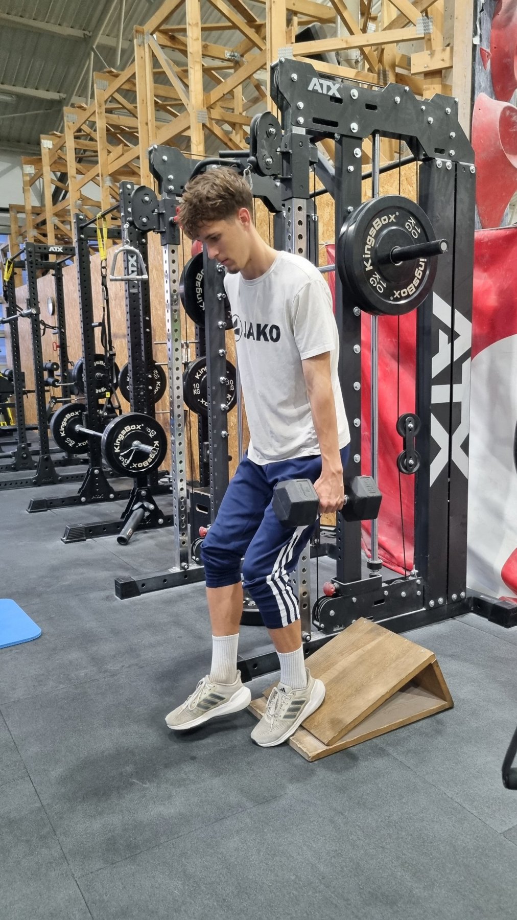
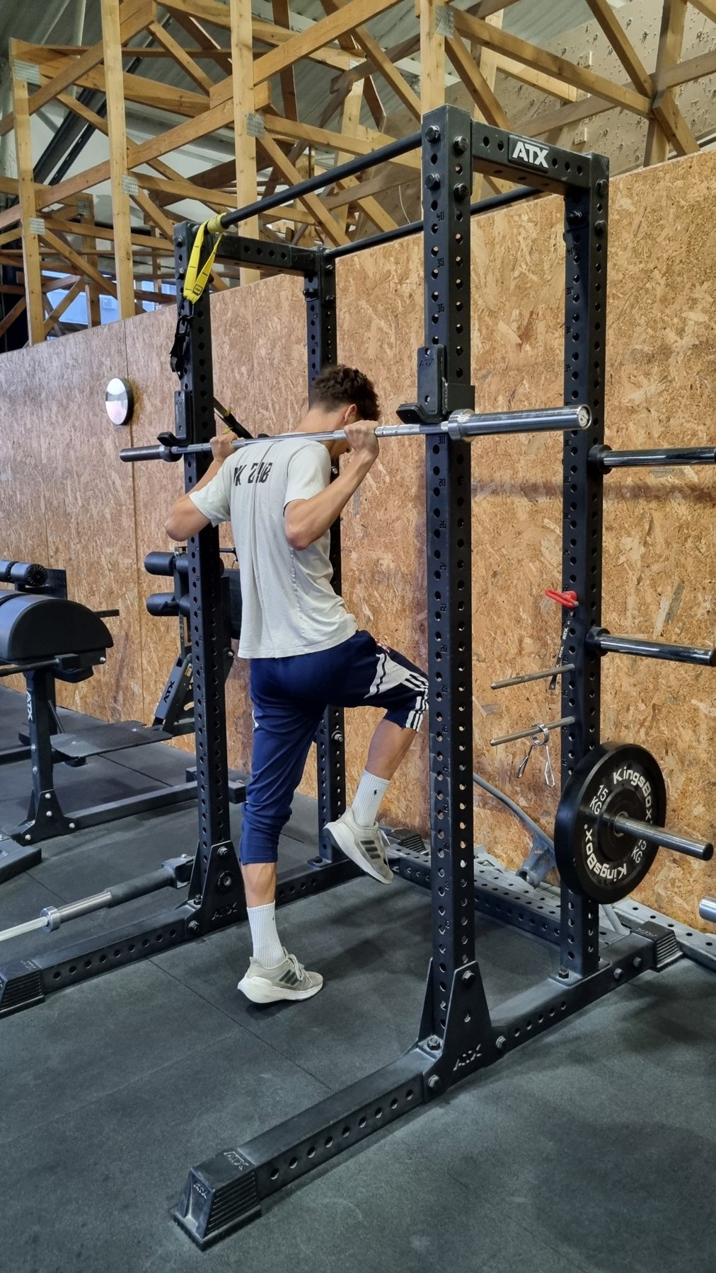
Isometric contractions come in two forms that are mechanically and neurally distinct (Schaefer & Bittmann, 2017). Yielding isometrics involve resisting an applied force while maintaining position, so the athlete holds under external load. Overcoming isometrics involve pushing against a fixed, immovable resistance, with no visible joint movement.
The clearest patellar-tendon-specific work on isometric loading comes from Rio et al. (2015). In athletes with patellar tendinopathy, a single session of heavy isometric contractions reduced tendon pain immediately, with the effect persisting for at least 45 minutes. The same session also increased maximal voluntary isometric contraction and reduced cortical inhibition, suggesting that the response involved both pain modulation and improved quadriceps drive.
This finding has not been consistently replicated. Later trials found that isometric and isotonic loading produced similar in-season pain relief (van Ark et al., 2016), and a randomised crossover trial found no clear analgesic advantage of isometric over dynamic loading (Holden et al., 2020). A systematic review concluded that isometric exercise is not superior to isotonic exercise for chronic tendinopathy and that the response to isometric exercise varies within and across tendinopathy populations (Clifford et al., 2020). In patients with patellar tendinopathy, a progressive tendon-loading programme that started with isometrics and progressed towards energy-storage tasks produced better clinical outcomes than an eccentric decline-squat programme (Breda et al., 2021). Isometric loading is therefore best viewed as one useful low-fatigue entry point that reduces pain and restores output in some athletes, rather than a reliable analgesic for everyone.
If quadriceps inhibition is part of the problem, restoring force output, not only managing pain, should be part of the approach. Heavy isometrics appear to address both in some athletes, and they allow high tendon force at a controlled joint angle without the loading rate of dynamic tasks.
Yielding isometrics are particularly practical in congested periods. After a match, the tendon has already been exposed to large volumes of sprinting, braking, cutting and landing. A yielding isometric provides a controlled mechanical input without adding further reactive or chaotic sport stimuli. It should not be interpreted as an immediate healing modality. The more accurate framing is that it can acutely reduce pain and restore quadriceps output when the tendon is sensitive, which makes it a useful low-speed loading option.
Direct evidence distinguishing overcoming from yielding isometrics for the patellar tendon is limited. In practice, overcoming isometrics are best framed as a high-force, low-movement option for situations where the goal is high voluntary drive and tendon tension without dynamic joint movement or eccentric braking demand.
Supramaximal eccentric loading
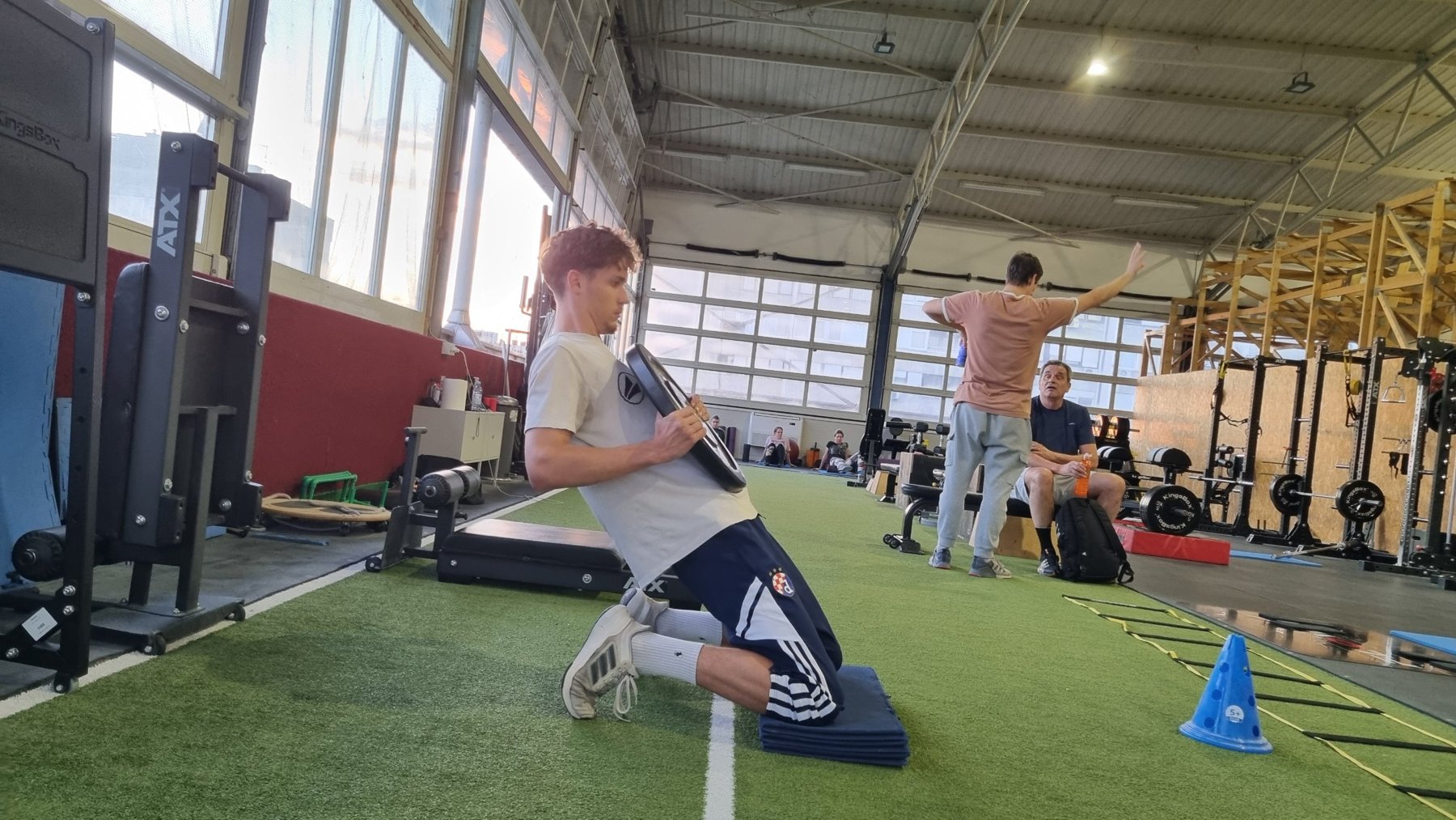
Supramaximal eccentric loading means lowering a load that is heavier than the athlete can lift, that is, more than 100% of concentric 1RM. This is possible because muscles are stronger when they lengthen than when they shorten, so an athlete can control a load in the eccentric phase that they could not lift in the concentric phase. The concentric phase is therefore assisted or removed, for example with weight releasers, a partner, flywheel or motorised devices, or a two-legs-up, one-leg-down technique (Wagle et al., 2017).
The rationale is that conventional training is limited by concentric strength, so the eccentric phase is always under-loaded. Supramaximal eccentrics close this gap and expose the quadriceps and patellar tendon to forces closer to those of braking and landing, where patellar tendon forces can exceed 7 BW (Verheul et al., 2024).
The cost is greater muscle damage, soreness and neuromuscular fatigue, especially in the first sessions. This cost falls quickly with repetition because of the repeated bout effect (Hyldahl et al., 2017), so supramaximal work should be introduced progressively and then kept in the programme regularly.
Peripheral adaptations
Peripheral adaptations are changes within the muscle and tendon themselves. For supramaximal and heavy eccentric training, the clearest changes are in muscle architecture and fibre phenotype, while gains in muscle size are similar to those produced by other forms of heavy loading.
Muscle architecture. Eccentric training lengthens muscle fascicles, which is generally interpreted as the addition of sarcomeres in series (Douglas et al., 2017). After 10 weeks of training in which the eccentric group lifted about 1.2 times the load of the concentric group, vastus lateralis fascicle length increased by 12% after eccentric training and by 5% after concentric training, whereas pennation angle increased by 5% and 30% respectively, with eccentric training favouring growth of the distal region of the muscle and activating MAPK signalling pathways that concentric loading did not (Franchi et al., 2014). In already-trained men, 10 weeks of accentuated eccentric loading with an eccentric load 40% above the concentric load increased resting fascicle length by about 14% in the vastus lateralis and 19% in the vastus medialis, whereas traditional training did not (Walker et al., 2020). Longer fascicles allow the muscle to produce force over a wider range of lengths and at higher shortening velocities, which is directly relevant to braking and landing, where the quadriceps works at long lengths under high load.
Muscle size and fibre type. Supramaximal eccentric loading does not appear to produce more hypertrophy than heavy conventional training. Accentuated eccentric loading and traditional training produced similar increases in muscle mass in trained men (Walker et al., 2016), neither protocol increased cross-sectional area over 9 weeks of elbow training with an eccentric load of about 120% of concentric 1RM (Brandenburg & Docherty, 2002), and a 2026 meta-analysis found no difference in muscle cross-sectional area between accentuated eccentric and constant-load training (Zhang et al., 2026). The fibre phenotype, however, may shift. In strength-trained athletes, 6 weeks of quadriceps training with eccentric overload increased the cross-sectional area of type IIX fibres, increased the proportion of type IIA fibres expressing MHC IIx mRNA and shifted gene expression towards a faster, more glycolytic profile, and only the eccentric overload group improved squat jump height (Friedmann-Bette et al., 2010).
Contractile properties. In one of the few truly supramaximal protocols, 4 weeks of plantar flexor training at 120% of concentric 1RM increased isometric torque by 30%, concentric torque by 14% and eccentric torque by 16%. Peak twitch torque increased from 14.6 to 17.4 N m and post-contraction potentiation increased by 18%, while the M-wave was unchanged. The authors nevertheless attributed most of the strength gain to neural factors, because the peripheral changes alone could not explain the increase in voluntary torque (Pensini et al., 2002).
Neural adaptations
The strongest argument for supramaximal eccentric loading is neural. The nervous system controls eccentric contractions differently from concentric and isometric contractions, and in untrained athletes it does not allow the muscle to use its full eccentric strength. This limitation is trainable, and it is most directly addressed by heavy eccentric loading. Because braking and landing are eccentric actions, this is where the transfer to sport is most direct.
Eccentric activation and neural adaptation. During maximal eccentric contractions the brain is highly active, but the spinal cord reduces the signal that reaches the muscle (Duchateau & Enoka, 2016). As a result, untrained individuals cannot fully activate their muscles when they lengthen under heavy load: in the quadriceps, the activation deficit has been reported as about 21% during eccentric contractions compared with 7% during isometric contractions (Beltman et al., 2004, as cited in Duchateau & Enoka, 2016). Highly trained athletes do not show this deficit (Amiridis et al., 1996), which indicates that it is trainable. Heavy resistance training increases eccentric strength together with muscle activation during eccentric contractions (Aagaard et al., 2000), and supramaximal eccentric training specifically increases the neural drive that reaches the muscle during eccentric contractions (Aagaard, 2018). After only 4 weeks of plantar flexor training at 120% of concentric 1RM, voluntary activation increased from 80% to 91% and co-activation of the opposing muscle decreased by 22% during eccentric contractions (Pensini et al., 2002). Eccentric training also reduces inhibition within the motor cortex (Kidgell et al., 2015; Colard et al., 2025) and transfers more strength to the untrained limb than concentric training does (Kidgell et al., 2015).
Relevance after injury. Because eccentric actions rely heavily on control from the brain, they can partly override the reflex inhibition of the quadriceps that follows knee injury (Stańczak et al., 2025). A similar effect may be relevant to the inhibited quadriceps in patellar tendinopathy (Rio et al., 2015), although this has not been tested directly.
Transfer to sport. In sport, braking and landing are not only tissue-capacity problems. They require the nervous system to produce and tolerate high force during lengthening, often under time pressure. Athletes with higher eccentric quadriceps strength generate greater horizontal braking forces (Stańczak et al., 2025), which links eccentric capacity directly to deceleration. Supramaximal eccentrics develop the capacity to control high force during lengthening in a way that isolated isometric work cannot.
On smaller screens, each row is shown as a labelled block.
Method | Key evidence | Use in the week |
|---|---|---|
Yielding isometrics Holding a position against an external load | In volleyball players with patellar tendinopathy, one session of heavy isometric quadriceps contractions reduced pain for at least 45 min (6.8 vs 2.6 out of 10 after isotonic loading), increased MVIC by 18.7% and reduced cortical inhibition (Rio et al., 2015). In-season, isometric and isotonic programmes reduced pain to a similar extent (van Ark et al., 2016), isometrics were not more analgesic than dynamic loading in a crossover trial (Holden et al., 2020), and the response varies within and across tendinopathy populations (Clifford et al., 2020). | MD+1 for controlled, low-fatigue tendon loading after the match; MD+2 to MD+3 above about 70% MVC alongside eccentric work; heavy holds above about 70% MVC on any day the tendon is painful or irritated |
Overcoming isometrics Pushing against an immovable resistance | Pushing and holding isometric actions differ mechanically and neuromuscularly (Schaefer & Bittmann, 2017). High tendon force can be produced without joint movement or eccentric braking. Heavy isometric contractions at 90% MVC, performed as 3 s contractions, increased Achilles tendon stiffness by 57% and Young's modulus by 51% over 14 weeks (Bohm et al., 2014), and a threshold above about 70% MVC appears practical for reaching high tendon strain (Merry et al., 2022). Direct patellar-tendon evidence is limited. | Throughout the week; on MD-1 and MD as part of the warm-up or in contrast methods paired with jumps or short sprints |
Supramaximal eccentrics Lowering a load above concentric 1RM with an assisted concentric phase | Peripheral: fascicle length increased by 12% after eccentric versus 5% after concentric training (Franchi et al., 2014) and by 14 to 19% with an eccentric load 40% above the concentric load in trained men (Walker et al., 2020); eccentric overload increased type IIX fibre size and fast-fibre gene expression (Friedmann-Bette et al., 2010); high-load eccentric training produced the largest gains in patellar tendon force, stiffness and modulus (Malliaras et al., 2013) and increased Achilles tendon stiffness by 82% and modulus by 86% (Geremia et al., 2018). Neural: 4 weeks at 120% of concentric 1RM increased voluntary activation from 80% to 91% and reduced co-activation of the opposing muscle by 22% (Pensini et al., 2002); supramaximal eccentrics increase neural drive during eccentric actions (Aagaard, 2018) and eccentric training reduces cortical inhibition (Kidgell et al., 2015; Colard et al., 2025). Performance: in trained men, accentuated eccentric loading increased isometric torque by 18% and eccentric torque by 10% (Walker et al., 2016). | MD+2 or MD+3 with a controlled eccentric phase of about 3 s, introduced progressively; reduced from MD-3 and minimal on MD-2 |
5. Practical integration
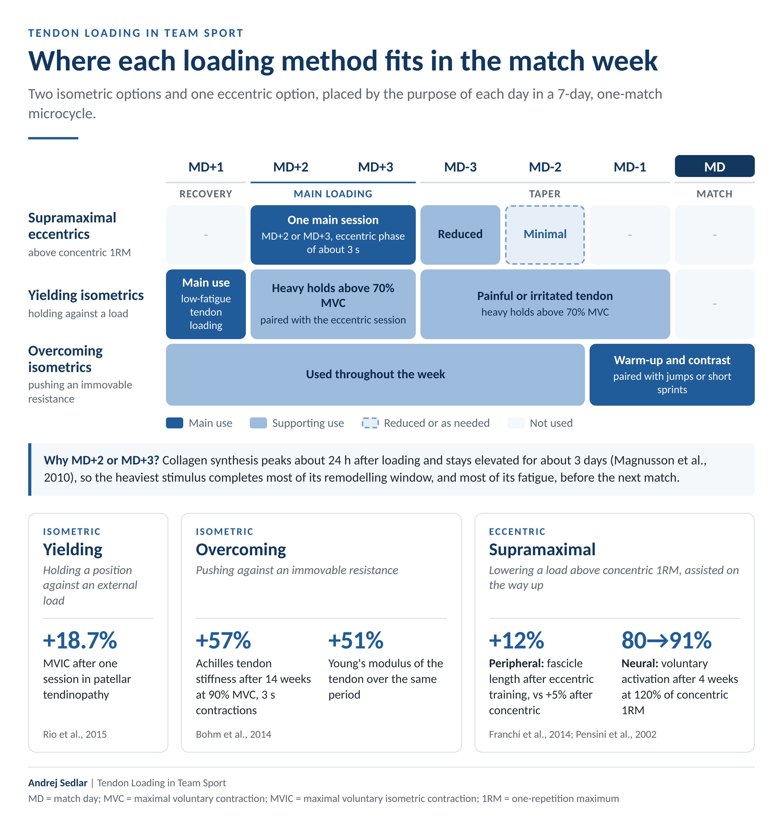
The practical question is not whether tendon loading should be included in the training week, but where it belongs within the weekly structure. In team sport, the match provides the highest sport-specific tendon exposure of the week through sprinting, braking, change of direction, jumping and landing. Additional tendon loading must therefore follow the logic of the microcycle rather than be added randomly.
The week can be understood through two connected layers. The first is the overall performance logic of the microcycle: match exposure, recovery, main loading days and a taper back into competition. The second translates that structure into tendon-loading decisions by distributing eccentric and isometric work according to the purpose of each training day.
Supramaximal eccentric loading is the most demanding option because it combines high force with muscle lengthening under load, so it belongs on MD+2 or MD+3. Collagen synthesis peaks around 24 hours after loading and remains elevated for about three days (Magnusson et al., 2010), so a heavy stimulus placed here completes most of its remodelling window, and most of the associated muscle fatigue, before the next match. A controlled eccentric phase of about 3 s matches the strain duration that produced the largest tendon adaptations (Bohm et al., 2014), and supramaximal work should be introduced progressively and then kept in the programme so that the repeated bout effect keeps its cost low (Hyldahl et al., 2017). From MD-3 onwards eccentric loading is reduced, and on MD-2 and MD-1 it is minimal or absent.
Isometric loading can be combined with eccentric work and used across the whole week, with the type of isometric chosen according to the day. Yielding isometrics suit MD+1, the day after the match, when the tendon has already been exposed to large volumes of sprinting, braking and landing. They load the tendon in a controlled, low-fatigue way while it recovers and can reduce pain and restore quadriceps output in some athletes (Rio et al., 2015), without adding further reactive load. On MD+2 and MD+3, heavy isometrics above roughly 70% MVC (Merry et al., 2022) can be paired with the eccentric session. Overcoming isometrics can be used throughout the week and are particularly useful on MD-1 and on match day itself, either as part of the warm-up or within contrast methods, where a short maximal overcoming isometric is paired with an explosive task such as a jump or a short sprint to prepare the athlete for competition.
Read the infographic as text
Where each loading method fits in the match week
Two isometric options and one eccentric option, placed by the purpose of each day in a 7-day, one-match microcycle.
MD+1: Recovery · MD+2 and MD+3: Main loading · MD-3, MD-2 and MD-1: Taper · MD: Match.
Supramaximal eccentrics
Above concentric 1RM.
- MD+1
- Not used.
- MD+2 or MD+3
- One main session. MD+2 or MD+3, eccentric phase of about 3 s.
- MD-3
- Reduced.
- MD-2
- Minimal.
- MD-1 and MD
- Not used.
Yielding isometrics
Holding against a load.
- MD+1
- Main use: low-fatigue tendon loading.
- MD+2 and MD+3
- Supporting use: heavy holds above 70% MVC, paired with the eccentric session.
- MD-3, MD-2 and MD-1
- Supporting use: painful or irritated tendon, heavy holds above 70% MVC.
- MD
- Not used.
Overcoming isometrics
Pushing an immovable resistance.
- MD+1, MD+2, MD+3, MD-3 and MD-2
- Supporting use: used throughout the week.
- MD-1 and MD
- Main use: warm-up and contrast, paired with jumps or short sprints.
Legend: Main use · Supporting use · Reduced or as needed · Not used.
Why MD+2 or MD+3? Collagen synthesis peaks about 24 h after loading and stays elevated for about 3 days (Magnusson et al., 2010), so the heaviest stimulus completes most of its remodelling window, and most of its fatigue, before the next match.
Isometric — Yielding
Holding a position against an external load.
+18.7% MVIC after one session in patellar tendinopathy.
Rio et al., 2015.
Isometric — Overcoming
Pushing against an immovable resistance.
+57% Achilles tendon stiffness after 14 weeks at 90% MVC, 3 s contractions.
+51% Young's modulus of the tendon over the same period.
Bohm et al., 2014.
Eccentric — Supramaximal
Lowering a load above concentric 1RM, assisted on the way up.
+12% Peripheral: fascicle length after eccentric training, vs +5% after concentric training.
80→91% Neural: voluntary activation after 4 weeks at 120% of concentric 1RM.
Franchi et al., 2014; Pensini et al., 2002.
Andrej Sedlar | Tendon Loading in Team Sport
MD = match day; MVC = maximal voluntary contraction; MVIC = maximal voluntary isometric contraction; 1RM = one-repetition maximum.
References
Tendon structure / mechanobiology
Heinemeier, K. M., Schjerling, P., Heinemeier, J., Magnusson, S. P., & Kjaer, M. (2013). Lack of tissue renewal in human adult Achilles tendon is revealed by nuclear bomb 14C. FASEB Journal, 27(5), 2074-2079.
Kjaer, M. (2004). Role of extracellular matrix in adaptation of tendon and skeletal muscle to mechanical loading. Physiological Reviews, 84(2), 649-698.
Magnusson, S. P., Langberg, H., & Kjaer, M. (2010). The pathogenesis of tendinopathy: Balancing the response to loading. Nature Reviews Rheumatology, 6(5), 262-268.
Screen, H. R. C., Birk, D. E., Kadler, K. E., Ramirez, F., & Young, M. F. (2015). Tendon functional extracellular matrix. Journal of Orthopaedic Research, 33(6), 793-799.
Zhang, S., Ju, W., Chen, X., Zhao, Y., Feng, L., Yin, Z., & Chen, X. (2022). Hierarchical ultrastructure: An overview of what is known about tendons and future perspective for tendon engineering. Bioactive Materials, 8, 124-139.
Tendon adaptation / loading
Arampatzis, A., Karamanidis, K., & Albracht, K. (2007). Adaptational responses of the human Achilles tendon by modulation of the applied cyclic strain magnitude. Journal of Experimental Biology, 210, 2743-2753.
Bohm, S., Mersmann, F., & Arampatzis, A. (2015). Human tendon adaptation in response to mechanical loading: A systematic review and meta-analysis. Sports Medicine - Open, 1, 7.
Bohm, S., Mersmann, F., Tettke, M., Kraft, M., & Arampatzis, A. (2014). Human Achilles tendon plasticity in response to cyclic strain: Effect of rate and duration. Journal of Experimental Biology, 217(22), 4010-4017.
Cook, J. L., & Purdam, C. R. (2009). Is tendon pathology a continuum? A pathology model to explain the clinical presentation of load-induced tendinopathy. British Journal of Sports Medicine, 43(6), 409-416.
Geremia, J. M., Baroni, B. M., Bobbert, M. F., Bini, R. R., Lanferdini, F. J., & Vaz, M. A. (2018). Effects of high loading by eccentric triceps surae training on Achilles tendon properties in humans. European Journal of Applied Physiology, 118(8), 1725-1736.
Merry, K., et al. (2022). Foundational principles and adaptation of the healthy and pathological Achilles tendon in response to resistance exercise. Journal of Clinical Medicine, 11(16), 4722.
Tendon forces / mechanical loading
Baxter, J. R., et al. (2021). Exercise progression to incrementally load the Achilles tendon. Medicine & Science in Sports & Exercise, 53(1), 124-130.
Garcia, S., Delattre, N., Berton, E., & Rao, G. (2022). Patellar tendon force differs depending on jump-landing tasks and estimation methods. Applied Sciences, 12(1), 488.
Verheul, J., Harper, D. J., & Robinson, M. A. (2024). Forces experienced at different levels of the musculoskeletal system during horizontal decelerations. Journal of Sports Sciences, 42(23), 2242-2253.
Patellar tendinopathy
Breda, S. J., et al. (2021). Effectiveness of progressive tendon-loading exercise therapy in patients with patellar tendinopathy. British Journal of Sports Medicine, 55(9), 501-509.
Clifford, C., et al. (2020). Effectiveness of isometric exercise in the management of tendinopathy: A systematic review and meta-analysis of randomised trials. BMJ Open Sport & Exercise Medicine, 6(1), e000760.
Holden, S., et al. (2020). Isometric exercise and pain in patellar tendinopathy: A randomized crossover trial. Journal of Science and Medicine in Sport, 23(3), 208-214.
Millar, N. L., et al. (2021). Tendinopathy. Nature Reviews Disease Primers, 7, 1.
Rio, E., et al. (2015). Isometric exercise induces analgesia and reduces inhibition in patellar tendinopathy. British Journal of Sports Medicine, 49(19), 1277-1283.
Tayfur, A., et al. (2022). Are landing patterns in jumping athletes associated with patellar tendinopathy? Sports Medicine, 52(1), 123-137.
van Ark, M., et al. (2016). Do isometric and isotonic exercise programs reduce pain in athletes with patellar tendinopathy in-season? A randomised clinical trial. Journal of Science and Medicine in Sport, 19(9), 702-706.
Wiesinger, H.-P., et al. (2020). Mechanical and material tendon properties in patients with proximal patellar tendinopathy. Frontiers in Physiology, 11, 704.
Loading methods / eccentric training
Brandenburg, J. P., & Docherty, D. (2002). The effects of accentuated eccentric loading on strength, muscle hypertrophy, and neural adaptations in trained individuals. Journal of Strength and Conditioning Research, 16(1), 25-32.
Douglas, J., Pearson, S., Ross, A., & McGuigan, M. (2017). Chronic adaptations to eccentric training: A systematic review. Sports Medicine, 47(5), 917-941.
Franchi, M. V., Atherton, P. J., Reeves, N. D., Flück, M., Williams, J., Mitchell, W. K., Selby, A., Beltran Valls, M. R., & Narici, M. V. (2014). Architectural, functional and molecular responses to concentric and eccentric loading in human skeletal muscle. Acta Physiologica, 210(3), 642-654.
Friedmann-Bette, B., et al. (2010). Effects of strength training with eccentric overload on muscle adaptation in male athletes. European Journal of Applied Physiology, 108(4), 821-836.
Hyldahl, R. D., Chen, T. C., & Nosaka, K. (2017). Mechanisms and mediators of the skeletal muscle repeated bout effect. Exercise and Sport Sciences Reviews, 45(1), 24-33.
Malliaras, P., et al. (2013). Patellar tendon adaptation in relation to load-intensity and contraction type. Journal of Biomechanics, 46(11), 1893-1899.
Pensini, M., Martin, A., & Maffiuletti, N. A. (2002). Central versus peripheral adaptations following eccentric resistance training. International Journal of Sports Medicine, 23(8), 567-574.
Schaefer, L. V., & Bittmann, F. N. (2017). Are there two forms of isometric muscle action? Results of the experimental study support a distinction between a holding and a pushing isometric muscle function. BMC Sports Science, Medicine and Rehabilitation, 9, 11.
Wagle, J. P., Taber, C. B., Cunanan, A. J., Bingham, G. E., Carroll, K. M., DeWeese, B. H., Sato, K., & Stone, M. H. (2017). Accentuated eccentric loading for training and performance: A review. Sports Medicine, 47(12), 2473-2495.
Walker, S., Blazevich, A. J., Haff, G. G., Tufano, J. J., Newton, R. U., & Häkkinen, K. (2016). Greater strength gains after training with accentuated eccentric than traditional isoinertial loads in already strength-trained men. Frontiers in Physiology, 7, 149.
Walker, S., Trezise, J., Haff, G. G., Newton, R. U., Häkkinen, K., & Blazevich, A. J. (2020). Increased fascicle length but not patellar tendon stiffness after accentuated eccentric-load strength training in already-trained men. European Journal of Applied Physiology, 120(11), 2371-2382.
Zhang, X., Weakley, J., Li, H., Marcos-Frutos, D., et al. (2026). Acute and chronic effects of accentuated eccentric loading vs. constant-load resistance training: A systematic review and meta-analysis. Sports Medicine, 56(7), 1749-1770.
Neural control of eccentric contractions
Aagaard, P. (2018). Spinal and supraspinal control of motor function during maximal eccentric muscle contraction: Effects of resistance training. Journal of Sport and Health Science, 7(3), 282-293.
Aagaard, P., Simonsen, E. B., Andersen, J. L., Magnusson, S. P., Halkjaer-Kristensen, J., & Dyhre-Poulsen, P. (2000). Neural inhibition during maximal eccentric and concentric quadriceps contraction: Effects of resistance training. Journal of Applied Physiology, 89(6), 2249-2257.
Amiridis, I. G., Martin, A., Morlon, B., Martin, L., Cometti, G., Pousson, M., & van Hoecke, J. (1996). Co-activation and tension-regulating phenomena during isokinetic knee extension in sedentary and highly skilled humans. European Journal of Applied Physiology, 73(1-2), 149-156.
Colard, J., Betus, Y., Tallio, T., Bizet, B., Nordez, A., Jubeau, M., & Cattagni, T. (2025). Eccentric training at long muscle lengths induces greater corticospinal and spinal reflex plasticity than eccentric training at short muscle lengths. Experimental Physiology. https://doi.org/10.1113/EP092470
Duchateau, J., & Enoka, R. M. (2016). Neural control of lengthening contractions. Journal of Experimental Biology, 219(2), 197-204.
Kidgell, D. J., et al. (2015). Increased cross-education of muscle strength and reduced corticospinal inhibition following eccentric strength training. Neuroscience, 300, 566-575.
Stańczak, M., Swinnen, B., Kacprzak, B., Pacek, A., & Surmacz, J. (2025). Neurophysiology of ACL injury. Orthopedic Reviews, 17, 129173. https://doi.org/10.52965/001c.129173
Biomechanics / deceleration
Harper, D. J., & Kiely, J. (2018). Damaging nature of decelerations: Do we adequately prepare players? BMJ Open Sport & Exercise Medicine, 4(1), e000379.
Komi, P. V. (2000). Stretch-shortening cycle: A powerful model to study normal and fatigued muscle. Journal of Biomechanics, 33(10), 1197-1206.
Lichtwark, G. A., & Wilson, A. M. (2007). Is Achilles tendon compliance optimised for maximum muscle efficiency during locomotion? Journal of Biomechanics, 40(8), 1768-1775.
CONTINUE LEARNING
Upcoming education and events
Loading dates…